Electrocardiogram
What can an ECG do?
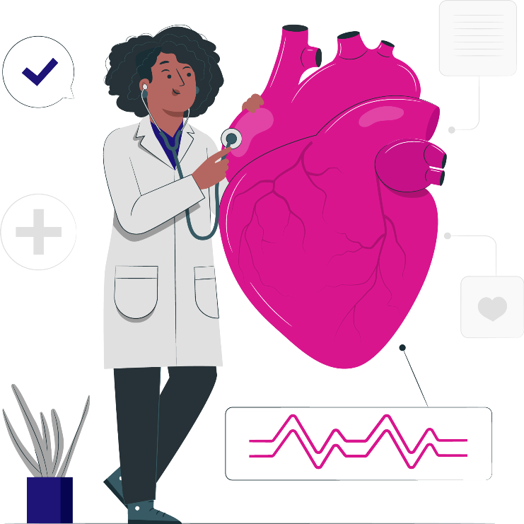
An electrocardiogram is a useful way to identify/detect damage in the heart or blood vessels caused due to high blood pressure.
Things an ECG can detect:
- Cholesterol clogging up on heart’s blood supply
- Heart Attack
- Enlargement in the heart
- Abnormal Heart Rhythms
How is an ECG carried out?
The ECG test takes only a few minutes. It is a safe and painless test that reads the signals of the heart and sends the readings to the electrocardiograph. The leads from an electrocardiograph machine are attached to the arms, legs, and chest using sticky patches. The machine then prints the information on a paper strip or on a monitor.

Types of ECG
Digital X-Ray
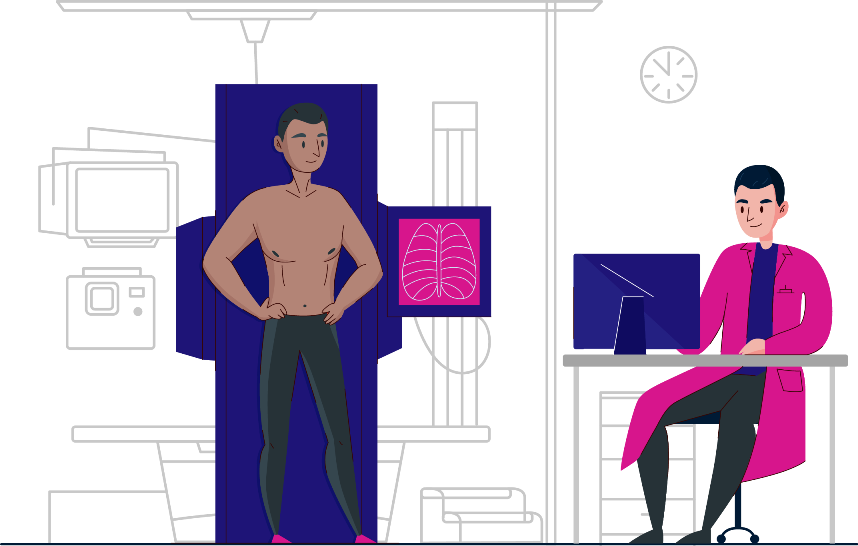
Digital radiography is a form of radiography that uses X-Ray sensitive plates to directly capture data, during the examination of the patient and immediately transfer it to a computer system without the use of an intermediate cassette. Instead of X-ray film, digital radiography uses a digital image capture device to capture data.


Ultrasound Imaging
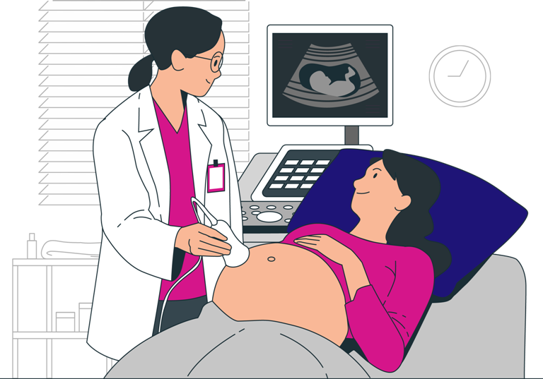
Ultrasound Imaging is a non-invasive medical examination that uses high-frequency sound waves to produce images of the internal organs of the body as well as the blood flowing through the blood vessels in real-time. This enables doctors to diagnose and treat a wide range of medical conditions.
Echocardiogram
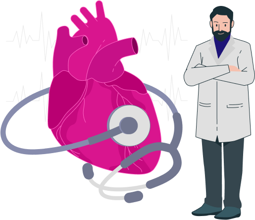
An echocardiogram uses sound waves to produce images of the heart. It allows doctors to check the heartbeat and its blood pumping potential along with heart diseases (if any).
Benefits of taking an echocardiogram test:
An echocardiogram helps in finding out the following:
- The shape, size, thickness, and movement of the heart's walls.
- The movement and the pumping potential of the heart.
- The workings of the valves of the heart.
- Identification of tumor, leakage of blood, or derangement around the heart valves.
- Problems with the outer lining and the large blood vessels of the heart.
- Blood clots and holes in the chambers of the heart.


